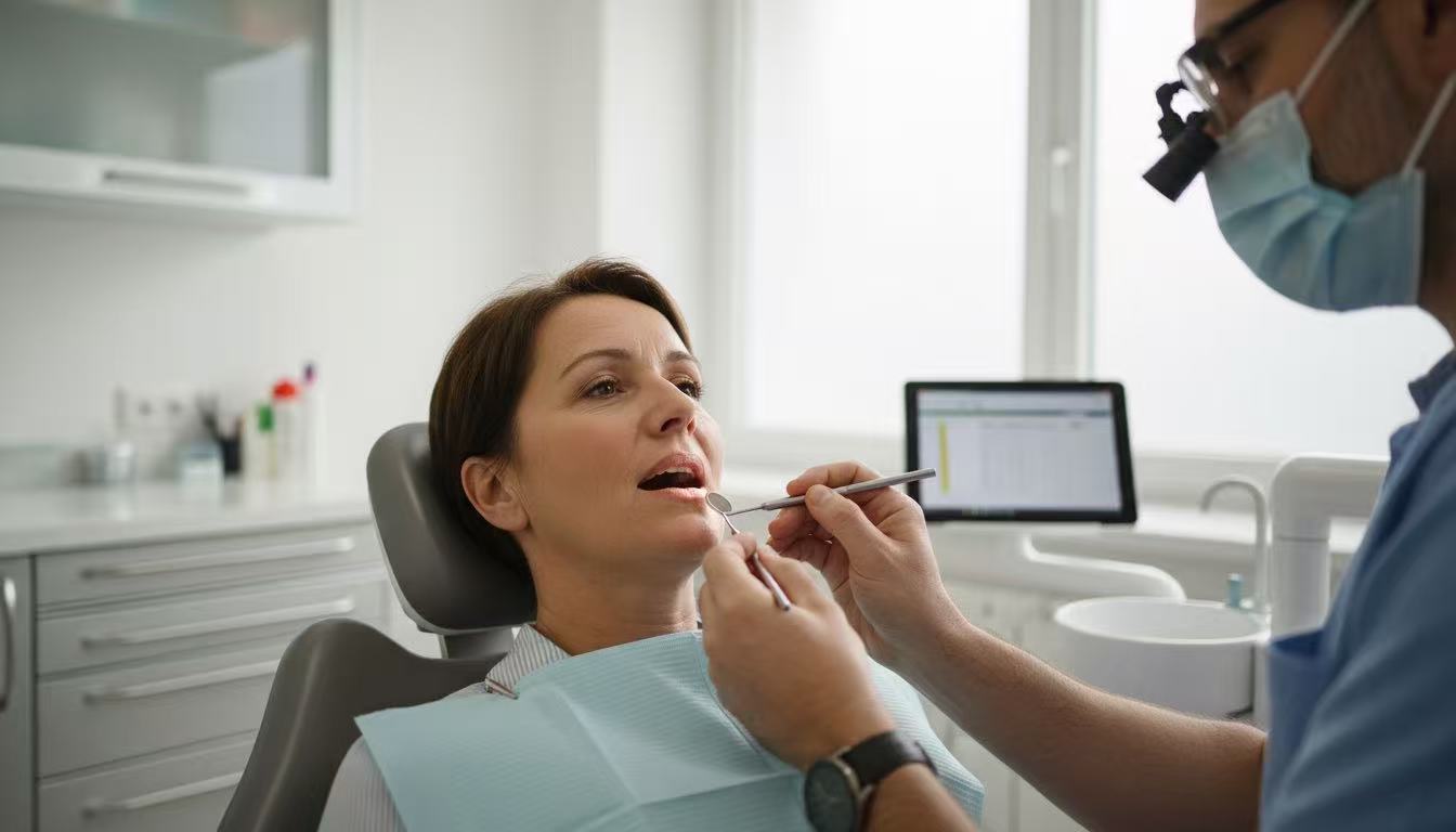









Tooth extraction often leads to alveolar bone resorption, which can compromise the structural integrity needed for future dental implants or restorations. Ridge preservation, a technique aimed at minimizing post-extraction bone loss, has gained prominence in modern dentistry. Among various techniques and materials, Platelet-Rich Fibrin (PRF) has emerged as a powerful and biocompatible tool. This article explores ridge preservation, focusing on the application of PRF to enhance outcomes.
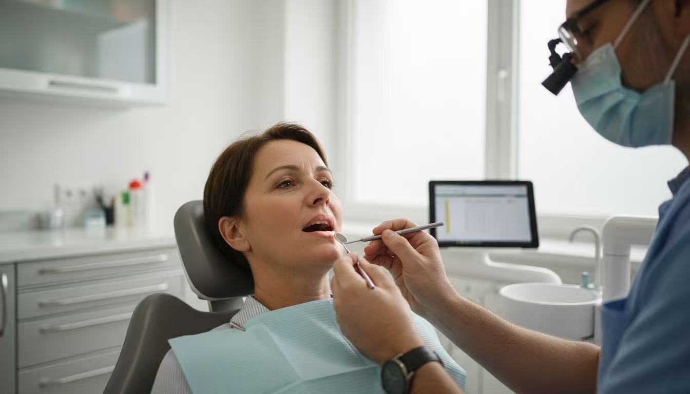
Parkway Dental Shanghai:The Importance of Ridge Preservation
Post-Extraction Bone Loss:
After tooth extraction, the alveolar bone undergoes remodeling, with significant resorption occurring in the first 3–6 months. This can lead to:
Reduced bone height and width.
Compromised aesthetics, especially in the anterior region.
Challenges in implant placement or prosthetic rehabilitation.
Ridge Preservation Goals:
The primary objective is to maintain bone volume and contour by preventing excessive resorption. This is achieved by filling the extraction socket with biomaterials that promote bone regeneration and soft tissue healing.
Techniques for Ridge Preservation
1. Grafting Materials
Autografts: Bone harvested from the patient, offering high biocompatibility.
Allografts: Bone from human donors, processed for safety.
Xenografts: Bone from animal sources, commonly bovine-derived.
Alloplasts: Synthetic materials like hydroxyapatite or calcium phosphate.
2. Membranes
Barrier membranes, either resorbable or non-resorbable, are used to protect graft materials and facilitate guided bone regeneration (GBR).
3. Advanced Biological Adjuncts
The use of biological agents, such as Platelet-Rich Fibrin (PRF), has revolutionized ridge preservation by enhancing natural healing processes.
What is PRF?
Definition:
Platelet-Rich Fibrin (PRF) is a second-generation platelet concentrate derived from the patient’s blood. Unlike Platelet-Rich Plasma (PRP), PRF is prepared without the use of anticoagulants or additives, resulting in a more natural, fibrin-rich matrix.
Composition:
PRF contains a high concentration of:
Platelets: Essential for clot formation and wound healing.
Growth Factors: Including vascular endothelial growth factor (VEGF), plateletderived growth factor (PDGF), and transforming growth factor-beta (TGF-β), which promote angiogenesis and tissue regeneration.
Leukocytes: Contribute to antimicrobial defense and healing.
Types of PRF:
L-PRF (Leukocyte-Rich PRF): High leukocyte concentration for enhanced antimicrobial properties.
A-PRF (Advanced PRF): Optimized for slower release of growth factors, extending the regenerative effect.

PRF in Ridge Preservation
Mechanism of Action
1. Bone Regeneration: PRF enhances osteoblast activity and bone formation through sustained release of growth factors.
2. Soft Tissue Healing: It accelerates epithelialization and reduces inflammation at the extraction site.
3. Angiogenesis: VEGF in PRF promotes new blood vessel formation, essential for nutrient delivery and tissue regeneration.
4. Antimicrobial Properties: Leukocytes in PRF help reduce infection risks postextraction.
Application of PRF in Ridge Preservation
1. Socket Filling: After tooth extraction, PRF membranes or plugs are placed directly into the socket. These act as scaffolds for tissue growth and clot stabilization.
2. Combination with Grafts: PRF can be mixed with bone graft materials to enhance handling and biological performance.
3. Membrane Replacement: PRF membranes can substitute for traditional barrier membranes, providing a resorbable, biocompatible option.
4. Flapless Techniques: PRF simplifies minimally invasive procedures, reducing patient discomfort and recovery time.
Advantages of PRF in Ridge Preservation
1. Biocompatibility: Autologous material eliminates risks of immune rejection or disease transmission.
2. Cost-Effectiveness: PRF preparation is relatively inexpensive compared to synthetic or donor-derived materials.
3. Ease of Use: Simple preparation protocol using a centrifuge machine.
4. Faster Healing: PRF accelerates soft tissue closure and minimizes postextraction complications.
5. Sustained Release: Growth factors are released over 7–14 days, promoting longterm regeneration.
Limitations of PRF
While PRF is a versatile and powerful adjunct, it has limitations:
• Volume Limitation: PRF alone may not provide sufficient bulk for larger defects. Combining it with graft materials is often necessary.
• Operator Dependency: Success depends on proper centrifugation protocols and handling.
• Short Working Time: PRF membranes need to be used quickly after preparation to retain their structure.
Conclusion
Ridge preservation is essential for maintaining alveolar bone integrity following tooth extraction, ensuring better outcomes for implants and restorations. PRF has become a valuable tool in this field, offering unparalleled advantages in enhancing bone regeneration and soft tissue healing. For dental professionals, understanding and incorporating PRF into clinical practice can significantly improve patient outcomes while aligning with modern, minimally invasive approaches.
About Parkway Dental Shanghai
Parkway Dentistry Center provide complete dental care for adults and children, all in one place. Our goal is simple: to help you keep your teeth healthy, comfortable, and looking good—now and in the long term.
We take time to explain findings, treatment options, costs, and timelines clearly, so patients can make informed decisions. Our team aims to provide a calm, respectful, and professional experience from the first visit through follow-up care.

References
1. Choukroun, J., et al. (2001). “Introducing Platelet-Rich Fibrin (PRF): A Second-Generation Platelet Concentrate.” Practical Procedures and Aesthetic Dentistry.
2. Miron, R. J., & Zhang, Y. F. (2018). “Autologous Platelet-Rich Fibrin: A Biological Scaffold and Healing Biomaterial.” Clinical Oral Implants Research.
3. Dohan Ehrenfest, D. M., et al. (2010). “Classification of Platelet Concentrates: From Pure Platelet-Rich Plasma (P-PRP) to Leukocyte- and Platelet-Rich Fibrin (L-PRF).” Trends in Biotechnology.
4. Goyal, L., et al. (2021). “Applications of PRF in Ridge Preservation and Implant Dentistry.” Journal of Oral Biology and Craniofacial Research.
5. Choukroun, J., et al. (2016). “Advanced PRF (A-PRF) in Dental Surgery:Extended Growth Factor Release and Benefits for Soft Tissue Healing.” European Journal of Oral Implantology.